AI acquires.
Physicians diagnose.
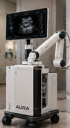
A Portuguese robotic ultrasound platform from the NWPT laboratory in Coimbra, built to bring standardised, diagnostic-quality examinations to every point of care, with or without a robot at the bedside.
Owning an ultrasound scanner is not ultrasound capacity
AURA targets the acquisition bottleneck. Acquisition becomes standardised and distributed, while interpretation stays with physicians and can remain centralised.
Operator-dependent
Good images need trained hands: probe positioning, contact pressure, the right acoustic window, parameter tuning and a judgement on whether the view is diagnostic. Skilled sonographers are scarce.
Unequal access
Primary-care units, regional facilities and mobile clinics often cannot staff an expert operator at all times. Patients travel, wait, or go without.
Variable quality
Exam quality depends on who holds the probe. An idle scanner when no expert is on shift is wasted capacity and delayed diagnosis.
One integrated medical-imaging product
A custom ultrasound probe and imaging engine, a force-controlled robotic arm, computer vision and clinical AI, co-designed under one design authority. It acquires standardised examinations without an expert sonographer manipulating the probe.
- Autonomous acquisition from patient detection to protocol completion
- AI anatomy recognition with real-time standard-view planning
- Closed-loop force control with gentle, compliant, validated limits
- Image-quality assessment: the system judges its own output
- Structured DICOM studies ready for PACS and physician review
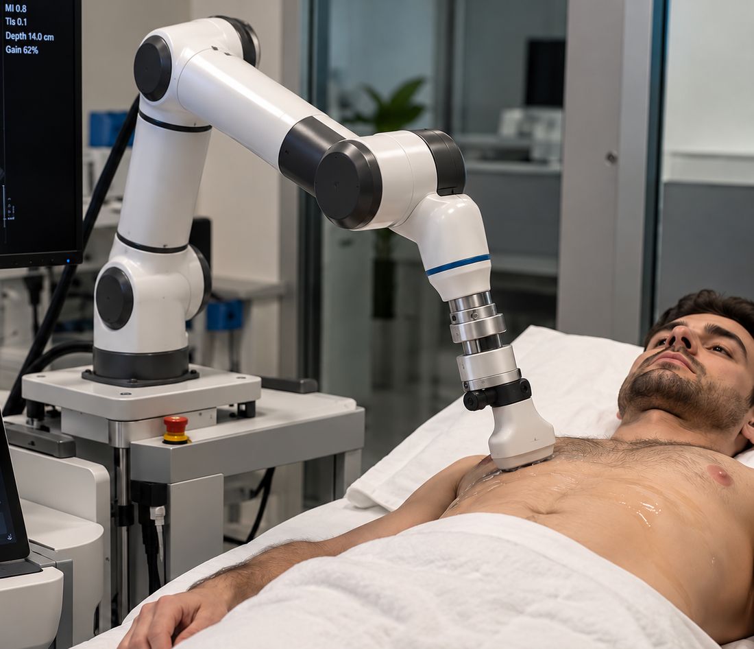
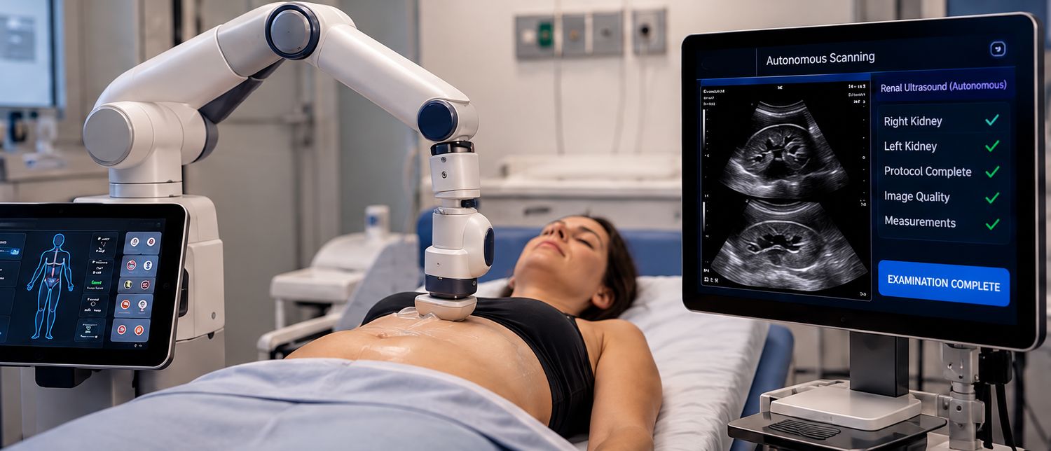
AURA at the bedside, as it would look in a ward
Concept visualizations of a human patient being examined in a clinical environment, with a live simulated ultrasound beside them. Move your mouse over the scene; the gold ring marks where the probe meets the patient and the live image follows the same renal protocol.
Illustrative waveform for atmosphere only. AURA is in engineering development and has no regulatory approval; nothing here is a clinical measurement.
Watch an exam, with the robot and without
A real-time 3D simulation of the renal protocol. Drag to rotate. Switch between the autonomous AURA robot and the handheld AURA probe, and follow the ultrasound plane as it finds the kidney.
- Position: vision finds the patient and target region
- Approach and contact under force limits
- Recognise anatomy: kidney and view plane
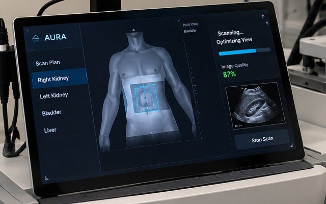
- Optimise position, tilt, force, depth, gain
- Assess quality and export DICOM
From patient to structured study, without a sonographer
Position
Vision identifies the patient, body orientation and target region.
Approach and contact
The robot moves under workspace, velocity and collision limits. SafeProbe establishes compliant contact.
Recognise anatomy
SonoAI detects the organ and estimates whether the plane matches the required clinical view.
Optimise in a loop
Navigator adjusts position, tilt and force while the imaging engine tunes depth, gain and focus.
Assess and complete
Quality is scored. Views, measurements and metadata are exported as DICOM.
Kidney views, simulated in real time and in 3D
AURA Renal is the first protocol: right kidney longitudinal and transverse, renal length, then the left kidney. Explore simulated B-mode, colour Doppler, reference views and an interactive 3D kidney.
Procedurally generated illustrations of renal echo-texture (capsule, cortex, medullary pyramids, central sinus). They are not patient scans and carry no diagnostic meaning.
Doppler on the AURA probe platform
Colour/PDI with steering, PRF, wall filter and invert; pulsed-wave and, on supported models, continuous-wave Doppler with baseline, angle and sample-volume controls.
- Colour ROI you can drag, resize and steer
- PW spectrum with adjustable sample volume
- Automatic blood-flow measurement on supported dual-head models
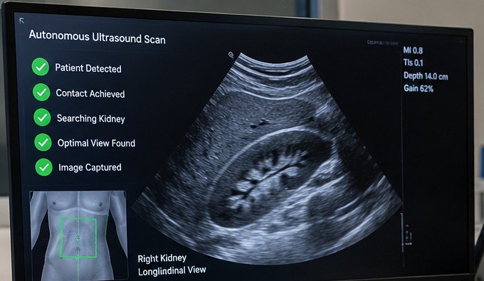
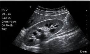
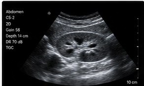
Reference renal views illustrate the standard outputs AURA Renal is designed to acquire. They are concept images, not patient data.
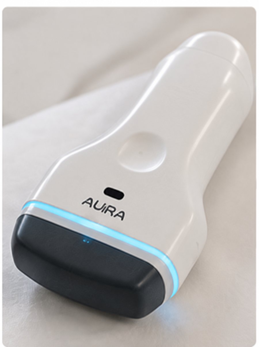
The AURA ultrasound probe platform
The same imaging core works as a wireless handheld probe. Connect over Wi-Fi or USB Type-C, scan on a tablet, phone or computer, and send studies to PACS.
*Availability of modes depends on the probe model and software release.
Inside the app
Tap a view to explore the interface.
AI proposes. Independent safety layers decide.
Safety control stays independent of the AI. Every autonomous action is logged from day one to support verification, design history and future regulatory evidence.
In-house design and integration
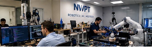
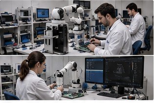
Portuguese architecture, IP, software, integration and verification, with a global supply chain for electronic components only.
AURA Probe
Mechanical design, housing, acoustic stack integration, assembly, cabling and calibration.
Ultrasound Engine
TX/RX board design, FPGA/GPU beamforming, reconstruction and real-time control firmware.
AURA Robotics
Arm kinematics and structure, joint control and motion planning.
SafeProbe
Compliant end-effector, force-control loop, safety mechanisms and emergency retraction.
Vision and compute
Patient localisation, calibration and perception software.
SonoAI and software
Anatomy recognition, Navigator, quality assessment, protocol engine, DICOM, cybersecurity and logging.
One platform, many organs
A common robotic and imaging core supports organ-specific packages as clinical evidence and regulatory scope expand. Acquisition at primary-care and mobile sites, interpretation by remote specialists.
Prototype for testing
Integrated laboratory prototype. Autonomous renal acquisition on phantom.
Test and refine
Phantom testing, image-quality tuning, force-control verification.
Complete integration
Integrate and refine probe, imaging electronics, robotics and software.
Full build completion
Integrated AURA Renal build, engineering verification and technical documentation.
Targets run from project kickoff and exclude licensing time. Human studies and clinical deployment follow ethics, safety and regulatory approvals. Planning targets assume funding, team availability and component supply.
Wider access, shorter waits, sovereign capability
Care
Wider access to ultrasound in underserved areas, shorter waiting times and fewer avoidable journeys, with consistent exam quality.
Specialists
Specialist time focused on interpretation and complex cases, with remote support through hub-and-spoke tele-ultrasound.
Industry
Sovereign capability in medical robotics, ultrasound engineering, embedded electronics and clinical AI, with high-value engineering jobs and an exportable MedTech platform.
Hospitals, partners and investors
We are looking for clinicians who can tell us where ultrasound access is hardest, technology partners, future distributors, and investors and funding programmes who share the vision.
- Clinical collaboration · feedback now, evaluation when prototype and approvals allow
- Distribution · after the regulatory steps are complete
- Investment and funding · introductory call and technical due diligence
Novea Wireless Power Transmission LDA
Coimbra, Portugal
research@nwpt.pt · www.nwpt.pt