
Operator-dependent
Diagnostic views need trained hands: pose, pressure, acoustic window, parameters. Without an expert on shift, the scanner sits idle.
AI acquires. Physicians diagnose.
AURA is a Portuguese autonomous ultrasound platform: a custom probe and imaging engine, a force-controlled robotic arm, computer vision and clinical AI in one product. It acquires standardised examinations without an expert sonographer holding the probe. Diagnosis stays with the physician.
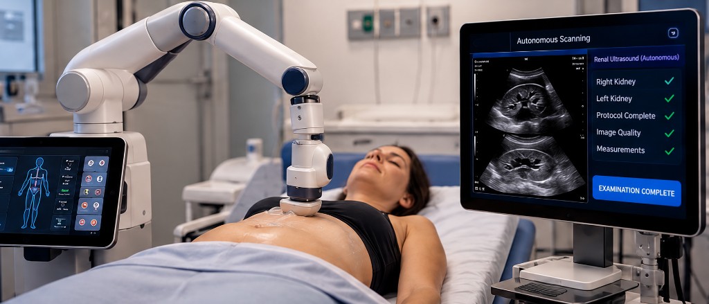
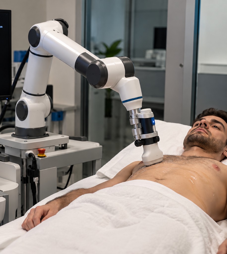
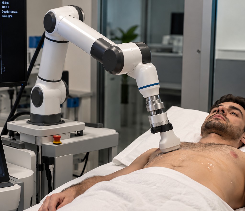
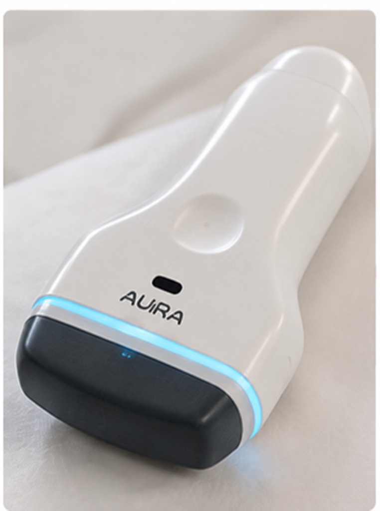
AURA does not replace the physician. This site does not claim MDR certification, CE marking, or measured clinical performance. Product figures are visualisations; final industrial design may differ. Orders and lead times: research@nwpt.pt.
Diagnostic views need trained hands: pose, pressure, acoustic window, parameters. Without an expert on shift, the scanner sits idle.
Primary-care, regional and mobile sites rarely staff a specialist operator at all times. Patients wait, travel, or go without.
Exam quality follows whoever holds the probe. AURA attacks the acquisition bottleneck: capture is standardised and distributed; interpretation can stay centralised.
One integrated product under a single Portuguese design authority — not a robot bolted onto a third-party scanner.
From patient detection to protocol completion.
Anatomy recognition and standard-view planning in real time.
Compliant contact, closed-loop force, validated limits.
The system judges its own output before storing it.
Renal first; bladder, thyroid, vascular and lung next.
A structured study with quality metadata for physician review.
Vision identifies the patient, body orientation and target region.
The robot moves under workspace, velocity and collision limits. SafeProbe establishes compliant contact.
SonoAI detects the organ and estimates whether the plane matches the required clinical view.
Navigator adjusts position, tilt and force while the imaging engine tunes depth, gain and focus.
Quality is scored. Views, measurements and metadata export as DICOM.
ACCEPT
The view meets the criteria and is stored.
RETRY
The view is inadequate; the system searches again.
ESCALATE
Persistent failure is handed to a human.
Ultrasound, robot pose, contact force and body vision feed a multimodal estimator. AURA does not merely move a probe or classify an image: it decides whether the exam has reached the acquisition endpoint.
Physical state (pose, force) and imaging state (depth, gain, focus, frequency) are tuned together.
A low-quality view is never silently counted as a completed exam.
AI proposes motion. The safety layer decides whether it is allowed.
Every autonomous action is logged. The design aims at ISO 13485 and EU MDR; this site does not claim certification or measured clinical performance.
Force, velocity, workspace, collision detection.
Watchdog, safe-state transitions, full action logging.
Hardware measurement off the AI path.
Passive compliance, hard stops, emergency stop, rapid probe retraction.
A clinically meaningful protocol with a bounded engineering and regulatory problem. It proves the full stack and opens the path to more organs.
In-house design and build. Electronics purchased. Portuguese architecture, IP, software, integration and verification.
Mechanics, housing, acoustic-stack integration, assembly and calibration. Transducer elements purchased.
TX/RX board, FPGA/GPU beamforming, real-time control firmware.
Kinematics, structure, motion planning and joint control.
Compliant end-effector, force loop, emergency retraction.
Patient localisation, calibration, perception, enclosure integration.
Anatomy, Navigator, quality, protocol engine, DICOM, cybersecurity and logging.
AURA does not replace the physician. This site does not claim MDR certification, CE marking, or measured clinical performance. Product figures are visualisations; final industrial design may differ. Orders and lead times: research@nwpt.pt.
Contact